Mostrando entradas con la etiqueta gross. Mostrar todas las entradas
Mostrando entradas con la etiqueta gross. Mostrar todas las entradas
jueves, 2 de junio de 2016
Embarazo molar (Mola hidatiforme completa)
La mola hidatidiforme es un embarazo que se caracteriza por una degeneración hidrópica de las vellosidades coriales y habitualmente la ausencia del feto. La mola parcial se caracteriza por ser resultado de una triploidía diándrica y por la presencia de cambios hidatiformes progresivos lentos con capilares vellosos funcionantes, que afectan solamente a algunas de las vellosidades; se asocia con un feto o embrión anormal identifcable (vivo o muerto), membranas o eritrocitos fetales.
[JUAREZ AZPILCUETA, Arturo; ISLAS DOMINGUEZ, Luis y DURAN PADILLA, Marco Antonio. MOLA HIDATIDIFORME PARCIAL CON FETO VIVO DEL SEGUNDO TRIMESTRE. Rev. chil. obstet. ginecol. [online]. 2010, vol.75, n.2 [citado 2016-06-02], pp. 137-139 . Disponible en: <http://www.scielo.cl/scielo.php?script=sci_arttext&pid=S0717-75262010000200011&lng=es&nrm=iso>. ISSN 0717-7526. http://dx.doi.org/10.4067/S0717-75262010000200011.]
martes, 15 de diciembre de 2015
Quiste de mesenterio (linfangioma)
Mesenteric cyst is defined as a cystic lesion located between the leaflets of the mesentery from the duodenum to the rectum , being most commonly found in ileum level. Since its first description in 1507 by Benevienae until 1993 there are only about 820 cases reported in the literature4 - 6.
Lymphangiomas are benign tumors, probably congenital, are more common in the cervical and axillary regions. They are unusual in abdominal and pancreas location. Its incidence is estimated at around 1:100,000 and 1:20,000 admissions in adults and in children. The first excision was performed by Tillaux (quoted Chung) only in 18025. Despite the long recognition of this disease, its origin classification and pathology remain controversial. The highest incidence is between the third and fourth decades of life, with 75% of those diagnosed after ten years with a slight female predominance. The term lymphangioma is appropriately used when there is hemodynamic isolation, or the injury is not related to the blood system10 - 13. Lymphangiomas are a major group of so-called vascular hamartomas, which result from a failure in the evolutionary development of the vascular system, including lymphatic and/or arteries and veins3.
[Tomado de: REIS, Diogo Gontijo Dos; RABELO, Nícollas Nunes and ARATAKE, Sidnei Jose.Mesenteric cyst: abdominal lymphangioma. ABCD, arq. bras. cir. dig. [online]. 2014, vol.27, n.2 [cited 2015-12-15], pp. 160-161 . Available from: <http://www.scielo.br/scielo.php?script=sci_arttext&pid=S0102-67202014000200160&lng=en&nrm=iso>. ISSN 0102-6720. http://dx.doi.org/10.1590/S0102-67202014000200016.]
Etiquetas:
aspecto,
gross,
linfangioma,
macro,
mesenterio,
pathology,
photo,
quiste,
tumor
viernes, 23 de octubre de 2015
Neurofibroma
Etiquetas:
foto,
gross,
macro,
micro,
nerve,
neurofibroma,
pathology,
patología,
photo,
sheat tumor
miércoles, 12 de agosto de 2015
Tumor miofibroblástico inflamatorio de colon
 | ||
Paciente de 4 años con tumor en colon transverso, el diagnóstico (por histología e inmunohistoquímica) fue tumor miofibroblástico inflamatorio
|
Independent of the location, IMTs are more common in children and young adults, but patients of any age and sex can be affected.1 Besides the lung, IMTs can also occur in the retroperitoneum, mediastinum, spleen, brain, pancreas, liver, or GI tract as single or multiple tumors.2,3,4,5,6,7 Although most authors admit that an ITM is a benign tumor, the recurrences and metastases presented in some of the reported cases1,6 may lead to a reclassification of this tumor as having uncertain malignant potential.
In the colorectal segment, the first case of IMT was described in the rectum by Coffin et al3 in 1995. Between 1995 and 2012, according to our knowledge, the number of reported cases rose to 24,1–17
Etiquetas:
colon,
de,
gross,
inflamatorio,
inflammatory,
macro,
miofibroblástico,
myofibroblastic,
pathology,
patología,
pseudotumor,
tumor
Miofibromatosis
 |
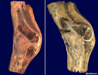
| Paciente con miofibromatosis, con paciente múltiples recurrencias y resecciones previas, se realizó amputación por dolor intratable. |
 |
| Aspecto histológico de los nódulos neoplásicos. |
Etiquetas:
amputación,
amputation,
dolor,
fibromatosis,
foto,
gross,
intratable,
macro,
miofibromatosis,
nodular,
pathology,
patología,
pieza,
tumor
Abscesos esplénicos piógenos / Spleen Abscesses (pyogenic)
 |
| Absceosos piógenos esplénicos. Paciente con inmunosupresión (leucemia linfoblástica tratada con quimioterapia) |
martes, 14 de julio de 2015
Quiste adrenal endotelial / Adrenal entothelial cyst
Etiquetas:
adrenal,
cyst,
endotelial,
entothelial,
foto,
gross,
imagen,
macro,
quiste,
simple,
suprarrenal,
vascular
jueves, 18 de junio de 2015
Secuestro pulmonar intralobar / intralobar pulmonary sequestration
Etiquetas:
foto,
gross,
imagen,
intralobar,
macro,
patología,
pulmonar,
pulmonary,
secuestro,
sequestration
Suscribirse a:
Entradas (Atom)


